 A Molecular Journey from DNA to RNA
1. Introduction
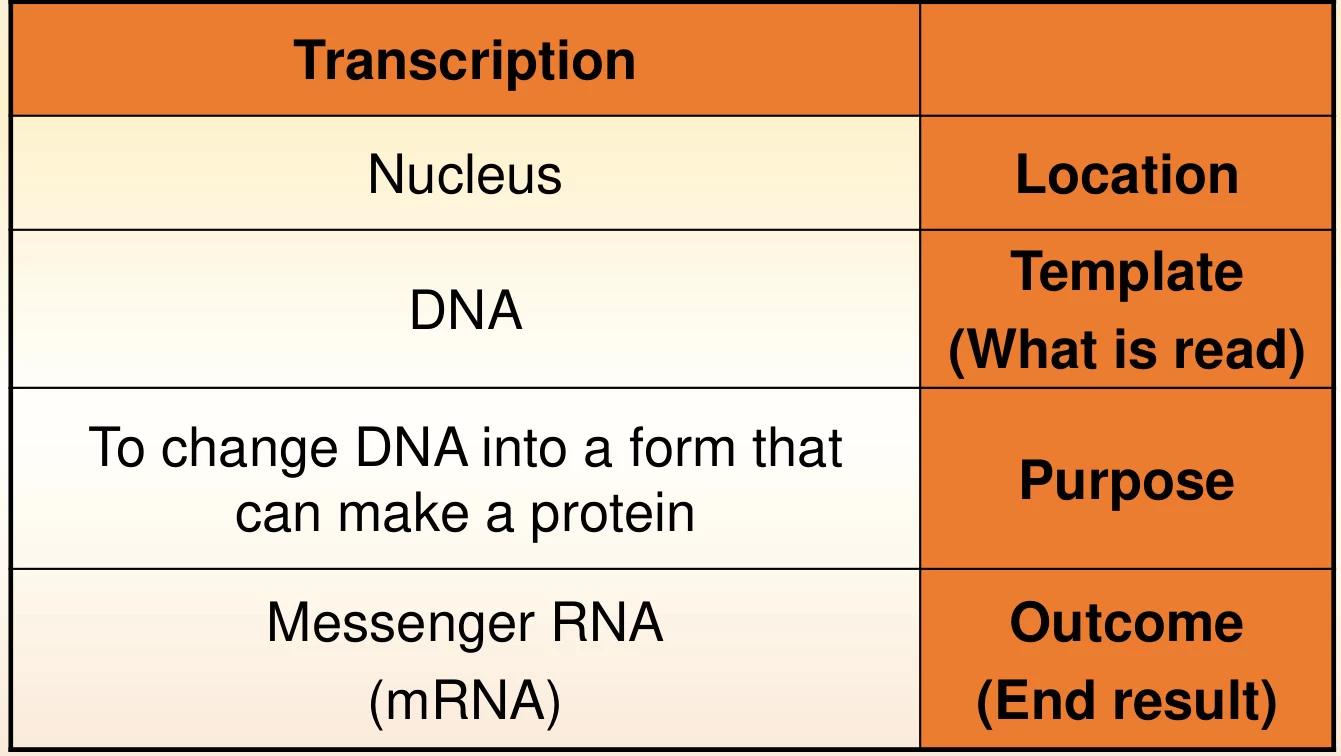
Transcription is the enzymatic process by which genetic information encoded in DNA is copied into a complementary RNA strand, serving as the first step in gene expression. This process, governed by RNA polymerase (RNAP), enables cells to translate genomic blueprints into functional molecules like mRNA, rRNA, and tRNA. Transcription occurs universally across prokaryotes and eukaryotes, though mechanistic and regulatory nuances distinguish these systems .
2. Structural Components: RNA Polymerase and DNA Template
RNAP is a multi-subunit enzyme that binds to specific DNA sequences called promoters to initiate transcription. In prokaryotes, RNAP consists of a core enzyme (α₂ββ’ω) and a dissociable sigma (σ) factor that directs promoter recognition. Eukaryotes utilize three specialized RNA polymerases:
- RNAP I: Synthesizes rRNA in the nucleolus.
- RNAP II: Produces mRNA and snRNA, regulated by phosphorylation of its C-terminal domain (CTD).
- RNAP III: Generates tRNA and 5S rRNA .
Suggested Figure: 3D model of RNAP bound to DNA, highlighting promoter regions and catalytic sites.
3. The Transcription Cycle
A. Initiation: Promoter Recognition and DNA Unwinding
Transcription begins when RNAP binds to promoter sequences. In prokaryotes, the σ factor guides RNAP to conserved motifs like the -35 hexamer and -10 Pribnow box. Eukaryotic RNAP II requires transcription factors (e.g., TFIID) to recognize the TATA box. DNA unwinding creates a transcription bubble (~14 base pairs), exposing the template strand for RNA synthesis .
Suggested Figure: Initiation complex showing RNAP, σ factor, and unwound DNA.
B. Elongation: RNA Synthesis
RNAP catalyzes the addition of ribonucleotides (NTPs) to the growing RNA chain in the 5’→3’ direction, guided by complementary base pairing (A-U, T-A, G-C). The enzyme moves processively along DNA, maintaining the transcription bubble while rewinding the upstream duplex. Unlike DNA polymerase, RNAP lacks proofreading, resulting in an error rate of ~10⁻⁴ .
Suggested Figure: Catalytic site of RNAP during elongation, illustrating NTP incorporation.
C. Termination: RNA Release
- Prokaryotes:
- Intrinsic termination: RNA hairpin structures followed by a poly-U tract destabilize the RNA-DNA hybrid.
- ρ-dependent termination: The ρ helicase binds nascent RNA, displacing RNAP .
- Eukaryotes: Termination involves polyadenylation signals (e.g., AAUAAA) and cofactors (CPSF, Pcf11) that cleave the transcript and recruit exonucleases .
Suggested Figure: Termination mechanisms: hairpin formation (prokaryotes) vs. polyadenylation (eukaryotes).
4. Post-Transcriptional Processing in Eukaryotes
Primary RNA transcripts undergo extensive modifications:
- 5’ Capping: A 7-methylguanosine cap is added to protect mRNA and aid ribosomal binding.
- Splicing: The spliceosome removes non-coding introns and joins exons, enabling alternative splicing to generate protein diversity.
- 3’ Polyadenylation: A poly(A) tail enhances stability and nuclear export .

Suggested Figure: mRNA processing: capping, splicing, and polyadenylation.
5. Prokaryotic vs. Eukaryotic Transcription
| Feature | Prokaryotes | Eukaryotes |
|---|---|---|
| Location | Cytoplasm | Nucleus |
| RNA Polymerase | Single multi-subunit RNAP | Three specialized RNAPs |
| Promoter Elements | -35/-10 motifs | TATA box, enhancers, silencers |
| Post-Processing | Minimal | Extensive (capping, splicing) |
| Regulation | Sigma factors, operons | Transcription factors, chromatin remodeling |
6. Biological and Technological Applications
- Biotechnology:
- mRNA vaccines (e.g., COVID-19) rely on in vitro transcription using phage RNAPs (e.g., T7 RNAP).
- CRISPR-Cas9 systems utilize RNAP-transcribed guide RNAs for genome editing .
- Medicine:
- Antibiotics like rifampicin inhibit bacterial RNAP to treat tuberculosis.
- Premature termination codon (PTC) therapies (e.g., ataluren) enable readthrough of faulty mRNA .
Suggested Figure: CRISPR-Cas9 system with RNAP-synthesized guide RNA.
7. Evolutionary Insights
RNAP’s core subunits (β, β’, α) are conserved across life domains, underscoring its fundamental role. Archaeal RNAP shares features with both eukaryotes and bacteria, offering clues to the evolution of transcriptional regulation .
8. Future Directions
- Structural biology: Cryo-EM and single-molecule studies are unraveling RNAP’s conformational dynamics.
- Synthetic biology: Engineered RNAPs with orthogonal promoter specificities enable advanced genetic circuits .
Data Source: Publicly available references.
Contact: chuanchuan810@gmail.com
