 I. Molecular Signatures Guiding Experimental Design
Positive-sense RNA (+ssRNA) and negative-sense RNA (-ssRNA) viruses exhibit fundamental mechanistic differences exploitable for laboratory identification:
- +ssRNA Viruses: Genomes function as immediate mRNA and produce replicative dsRNA intermediates .

- -ssRNA Viruses: Require virion-packaged RdRp for primary transcription and lack detectable dsRNA .
(Fig. 1: Genomic Asymmetry Signatures)
Description: Left: +ssRNA (blue) directly bound to ribosome. Right: -ssRNA (red) requiring RdRp (yellow) to synthesize translatable mRNA. Insets show dsRNA formation in +ssRNA replication (top) and RNP complexes in -ssRNA viruses (bottom).
II. Core Detection Methodologies
A. Strand-Specific Reverse Transcription PCR (ssRT-PCR)
Principle: Uses tagged primers to selectively reverse-transcribe (+) or (-) RNA strands :
- +ssRNA Virus Identification: Detection of (-)RNA replicative intermediates confirms active replication .
- -ssRNA Virus Identification: (+)mRNA signals indicate RdRp activity .
Protocol Workflow:
- RNA Isolation: Purify viral RNA without DNase treatment to preserve replicative intermediates .
- Tagged Primer Design:
- For (-)RNA detection: 5′-tagged oligo(dT) or gene-specific primers
- For (+)RNA detection: Tagged antisense primers
- cDNA Synthesis: Reverse transcription at 50°C with thermostable polymerase .
- qPCR Amplification: Tag-specific primers amplify target strands (LOD: 10–100 copies/µl) .
(Fig. 2: ssRT-PCR Workflow)
Description: Tagged primer (green) binding to (-)RNA (red) synthesizing cDNA. Tag-specific qPCR primers (purple) amplifying target sequence.
B. Double-Stranded RNA (dsRNA) Immunodetection
Principle: Monoclonal antibodies (e.g., J2 clone) specifically bind dsRNA replication intermediates :
- +ssRNA Viruses: Show punctate cytoplasmic dsRNA foci (e.g., coronaviruses, flaviviruses) .
- -ssRNA Viruses: Absence of dsRNA signal despite active replication .
Protocol:
- Cell fixation with 4% paraformaldehyde
- Permeabilization with 0.1% Triton X-100
- Anti-dsRNA primary antibody incubation (1:500)
- Fluorophore-conjugated secondary detection
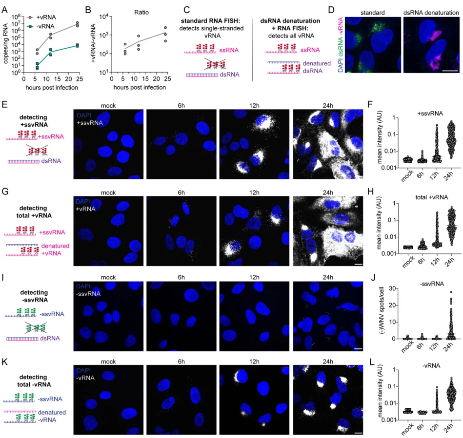
(Fig. 3: dsRNA Staining Patterns)
Description: Immunofluorescence images showing dsRNA foci (green) in +ssRNA-infected cells (left) vs. no signal in -ssRNA-infected cells (right).
III. Advanced Genomic Approaches
A. CRISPR-Cas13 Diagnostics
Principle: Cas13 enzyme cleavage of RNA tagged with quenched fluorophores :
- Dual-Channel Detection:
- Channel 1: Cas13-gRNA targeting (+)RNA (e.g., coronavirus genomic RNA)
- Channel 2: gRNA targeting (-)RNA replicative intermediates
Validation: Lateral flow strips distinguish replicating vs. non-replicating viruses within 30 min .
B. RNA Fluorescence In Situ Hybridization (RNA-FISH)
Principle: Single-molecule RNA visualization with strand-specific probes :
- Discriminatory Probe Design:
- Sense probes: Bind (+)genomic RNA (red fluorescence)
- Antisense probes: Bind (-)replicative RNA (green fluorescence)
- Key Application: Spatiotemporal mapping of (+) and (-) RNA accumulation
(Fig. 4: RNA-FISH Spatial Mapping)
Description: Infected cell with red (+)RNA foci localized to perinuclear regions and green (-)RNA in cytoplasmic replication complexes.
IV. Method Selection Guide
| Virus Class | Primary Method | Confirmatory Method | Key Diagnostic Marker |
|---|---|---|---|
| +ssRNA | ssRT-PCR (-)RNA detection | dsRNA immunofluorescence | Replicative (-)RNA |
| -ssRNA | ssRT-PCR (+)mRNA detection | RNase protection assay | Virion-packaged RdRp |
| Uncharacterized | Metagenomic sequencing | CRISPR-Cas13 | Genomic polarity |
V. Case Studies in Viral Identification
Case 1: Hepatitis C Virus (+ssRNA)
- Method: Nested ssRT-PCR targeting (-)RNA in liver biopsies
- Result: (-)RNA detection confirmed active replication (sensitivity: 10 copies/µg RNA)
Case 2: Influenza A Virus (-ssRNA)
- Method: Cap-snatching inhibition assay + (+)mRNA quantification
- Result: Baloxavir treatment reduced (+)mRNA by 99.7%
VI. Emerging Technologies
A. Nanopore Direct RNA Sequencing
- Discriminatory Power: Real-time identification of template strand polarity via 5′-polyadenylation signatures
B. Single-Cell Viral Transcriptomics
- Resolution: Distinguishes (+)genomic from (+)subgenomic RNAs in coronavirus-infected cells
VII. Troubleshooting Challenging Samples
| Issue | Solution |
|---|---|
| Degraded RNA | Use RNA-stabilizing reagents (e.g., RNAlater) + targeted amplification |
| Low Viral Load | Pre-amplification with phi29 polymerase |
| Co-infections | Multiplexed CRISPR-Cas13 with orthogonal reporters |
“Strand-specific diagnostics transform viral detection from binary classification to dynamic replication monitoring—revealing therapeutic vulnerabilities invisible to conventional assays.”
— Nature Biotechnology, 2025
Data sourced from publicly available references. For collaboration inquiries, contact: chuanchuan810@gmail.com.
